Committed to Eye Care with Compassion, Technology and Competency

Mukundapur: 844-480-0008
VIP Road: 033 2570 0097
Committed to Eye Care with Compassion, Technology and Competency
Home / State of the Art Equipment
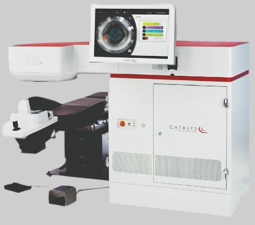
The Femto Laser Cataract Surgery machine offers a precision-guided approach to cataract removal, using femtosecond laser technology. This machine ensures a highly accurate, minimally invasive procedure, improving safety and recovery times. It replaces traditional manual methods, reducing the risk of complications and enhancing visual outcomes, particularly for complex cases. This state-of-the-art technology delivers exceptional precision and is a key innovation in modern cataract treatment.
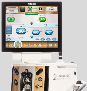
The Centurion® Vision System is the central part of the Cataract Refractive Suite by Alcon. It is an adaptive phaco technology that automatically and continuously adjusts to the changing conditions within the eye intra-operative. The new wireless footswitch optimises safety in the operating room. This integrated, automated system intelligently optimises chamber stability, allowing surgeons to safely perform a perfect optimal phacoemulsification.
The Verion™ technology is an innovative, accurate imaging system used to help customize the cataract procedure process and provide more precise lens placement. This revolutionary technology creates a detailed map of the eye's unique features before cataract surgery. It is designed to avoid the plethora of errors that could potentially occur along the surgical course, beginning with the initial evaluation through to the execution of the procedure.
A high-resolution image is taken of the patient's eye, mapping out important details and measurements. This map allows the surgeon to more efficiently plan, and customize, each step of the procedure to the patient's eye. The data received is transferred to display in the microscope in the operating room. By integrating the planning phase of the cataract surgery with the surgery itself, the system creates improvements in accuracy, precision, and safety throughout the duration of the process.
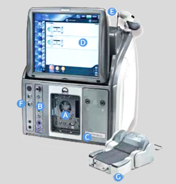
The Alcon CONSTELLATION Vision System is an ophthalmic microsurgical system that is indicated for both anterior segment (phacoemulsification & removal of cataracts) & posterior segments (Vitreoretinal) ophthalmic surgery. This machine’s user-friendly interface, advancements in high speed cutting, intraocular pressure control, illumination, laser and other features allows for more surgeon control during retinal surgery and makes it a a game changer in vitreoretinal surgery.
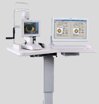
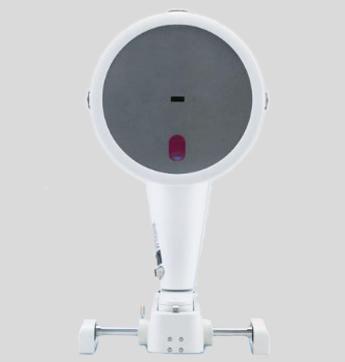
Heidelberg Retinal Angiogram working on principle of confocal scanning laser imaging provides a high resolution of retinal and choroidal vasculature with low light exposure providing comfort and safety for the patient.
Enhanced contrast, details and image sharpness are generated using confocality. Simultaneous digital FA and ICGA images with three-dimensional resolution offer improved diagnosis of retinal and choroidal pathologies. The machine offers advantage of high resolution and high contrast, low light exposure, simultaneous FA and ICGA imaging, fundus autoflurosense, dynamic high speed angiography, wide field imaging and composite imaging and iris imaging. It also provides non-invasive techniques like blue reflectance, FAF and infrared imaging.
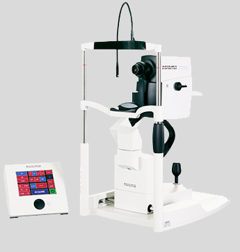
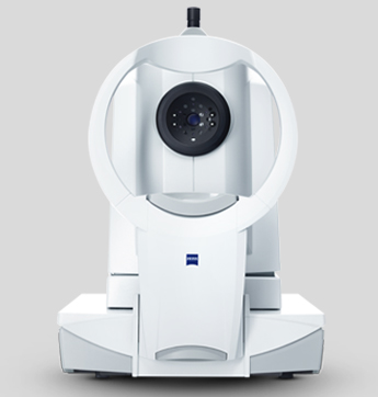
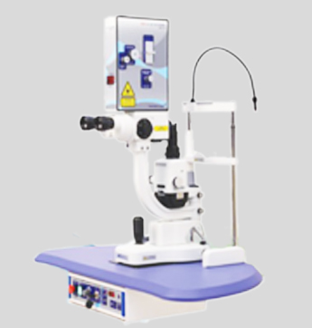
This machine from Zeiss is an impressive, powerful, diode-pumped solid-state laser for controlled and gentle photocoagulation of the retina. It has sufficient power reserves for any treatment strategy – whether it is with short laser pulses in the millisecond range for a gentle grid treatment, or long pulses for an effective retinopexy or even angioma sclerotherapy. Its built-in thermoelectric cooling system ensures maximum temporal stability of the laser power and thus meets the basic prerequisite for reproducible clinical results. It delivers a homogeneous, sharply-defined and reproducible laser spot on the retina, which minimizes heat-related side effects on the patient’s cornea. The active ClearView physician safety filter offers not only a unique and true-to-color slit lamp image, but also reliably protects the physician, automatically swinging into position when the therapy beam is activated.
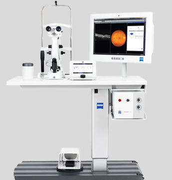
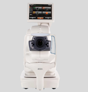
The IOLMaster 700, component of the ZEISS Cataract Suite, is the latest technology which provides highly repeatable and accurate measurements for lens power calculation before cataract surgery. It is non-invasive, very fast one machine stop for cataract surgery work up which gives impressive results in all types of cataracts including post refractive surgery.
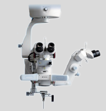
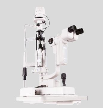
It is the new standard in versatile microsurgery. The new and innovative design allows the surgeon to view a sharper image, with more clarity than ever before and provides a natural stereoscopic view making it a perfect aid for your surgeon to work from a comfortable working distance. The built in yellow filter provides full protection against retina phototoxicity making it a safe choice.
Optical coherence tomography angiography (OCT-A) is a non-invasive technique for imaging the microvasculature of the retina and the choroid. OCT-A technology uses laser light reflectance of the surface of moving red blood cells to accurately depict vessels through different segmented areas of the eye, thus eliminating the need for intravascular dyes. With main advantages of shorter acquisition time and being non-invasive, it has become the latest technology in diagnosis and follow up of many retinal diseases, perfusion analysis in glaucoma and also uveitis.
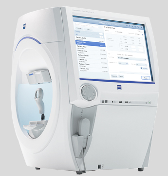
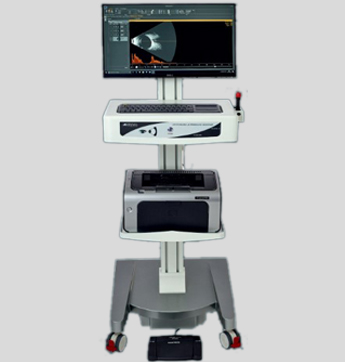
The primary function of the visual field test is to measure the patient’s peripheral and central vision with repeated light stimulus. Humphrey Visual Field Analyser is a state of the art technology used for screening, monitoring and assisting in the diagnosis of glaucoma and certain neuro-ophthalmological conditions. It is a non-invasive technique which offers different strategies and protocols to suit different patients needs while providing your doctor with the information required. The results are compared with those of the age-matched normal population as well as tracking each individual patient's possible changes over time, thus helping in monitoring progression of the disease and efficacy of treatment.
Pentacam is the Gold Standard in anterior eye segment tomography by OCULUS, Germany. It is a Comprehensive Anterior Segment Analyser providing in about 2 seconds Scheimpflug image of anterior segment, 3D anterior chamber analysis, pachymetry, corneal topography and cataract analysis. So in very less time, one machine provides organised information at a glance required for diagnosis of different anterior segment diseases and work up for refractive surgeries thus helping your surgeon in better planning and delivering excellent results.
The ultrasound scan machine offers two-dimensional cross-sectional view of the eye as well as the orbit. It offers a quick, non-invasive test to assess the structural integrity and pathology of the eye. It can provide additional information not readily obtained by direct visualization of ocular tissues and is particularly useful in patients with pathology that prevents or obscures ophthalmoscopy (e.g., large corneal opacities, dense cataracts, or vitreous haemorrhage).
A common problem post cataract surgery is clouding of the membrane known as the bag in which the artificial lens is placed. This change referred to as posterior capsular opacification can occur soon after surgery or years later and affects the clarity of vision as light rays don’t reach the posterior part of the eye.
Yag Capsulotomy is the easy solution to this problem. The Yag Cap machine uses laser beams to clear the membrane in a matter of minutes in an out patient setting. It is an easy, safe and painless procedure which improves vision quickly.
Ultrasound biomicroscopy (UBM) is a high-resolution ultrasound technique that allows noninvasive in vivo imaging of structural details of the anterior segment of the eye at near light microscopic resolution and provides detailed assessment of anterior segment structures, including those obscured by normal anatomic and pathologic relations. This machine is useful in providing important information in diagnosis and management of lid, corneal, ocular surface ,anterior chamber pathologies and glaucoma. It is a non-invasive and painless investigation.
Selective Laser Trabeculoplasty is a laser treatment for open-angle glaucoma that lowers eye pressure. It can be used as initial treatment, instead of eye drop medications, or as additional treatment when medications do not adequately reduce the eye pressure. The machine allows to apply laser energy to drainage tissue in the eye. This starts a chemical and biological change in the tissue that results in better drainage of fluid through the drain and out of the eye eventually resulting in lowering of IOP. This type of laser used has minimal heat energy absorption because it is only taken up by selected pigmented tissue in the eye, hense the name selective, thus producing less scar tissue and minimal pain. This machine allows for a safe, quick and pain free in office procedure for management of glaucoma.
Slit lamp evaluation is a part of basic ophthalmology work up. Slit lamp photography allows for documentation of the same providing magnified photographs with diffuse or slit light focusing on the required structure to highlight pathology. This helps to maintain record for the disease and also check for progression or regression helping in better management of disease.
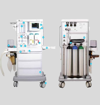
The GE Datex Ohmeda is an advanced unit for general anaesthesia. This is a fail safe device which ensures that whenever oxygen pressure is reduced and until flow ceases, the set oxygen concentration shall not decrease at the common gas outlet thus making it a safe device. Equipped with high priority visible and audible alarms makes it an excellent choice for general anaesthesia.